
Cone Beam Computed Tomography (CBCT) is an advanced three dimensional dental imaging technology that enables endodontists to examine teeth, root canals, surrounding bone, and nearby anatomical structures with exceptional accuracy. Compared to traditional two dimensional dental X-rays, CBCT provides a comprehensive 3D view that supports more accurate diagnosis, improved treatment planning, and more predictable outcomes during modern endodontic care.
Modern Technology Is Changing the Future of Endodontics
Technology continues to transform every area of healthcare, and dentistry is no exception. One of the most significant advancements in modern endodontics is Cone Beam Computed Tomography, more commonly known as CBCT. This sophisticated imaging technology has changed how endodontists diagnose complex dental conditions, plan treatment, and preserve natural teeth.
Endodontics focuses on diagnosing and treating diseases that affect the inside of the tooth, including the dental pulp and root canal system. Because many of these conditions develop beneath the surface of the tooth and are hidden from direct view, accurate diagnosis is one of the most important factors in successful treatment. Even small anatomical variations or hidden infections can influence treatment decisions and long term outcomes.
Traditional dental radiographs remain valuable diagnostic tools, but they produce only a two dimensional image of a three dimensional structure. While they continue to play an important role in everyday dentistry, certain cases require more detailed visualization. Cone Beam Computed Tomography provides that additional level of detail by producing highly accurate three dimensional images that allow endodontists to evaluate the tooth and surrounding structures from virtually every angle.
At Delta Root Canal, advanced diagnostic technology supports careful treatment planning and individualized patient care. Using modern imaging systems when clinically appropriate allows treatment decisions to be based on comprehensive information rather than assumptions, helping preserve natural teeth whenever possible.
What Is Cone Beam Computed Tomography (CBCT)?
Cone Beam Computed Tomography is a specialized form of dental imaging that produces highly detailed three dimensional images of the teeth, jaw, surrounding bone, nerves, and other oral structures. Unlike conventional dental X-rays that capture only a flat image, CBCT creates a complete digital reconstruction of the treatment area.
During the scan, a cone shaped X-ray beam rotates around the patient’s head while capturing hundreds of individual images from multiple angles. Specialized computer software then combines these images into a highly detailed three dimensional model that can be viewed from different perspectives.
This technology gives endodontists an exceptional view of the internal anatomy of each tooth, making it easier to identify conditions that may not be visible on traditional radiographs.
CBCT imaging can help evaluate:
- Root canal anatomy
- Additional or hidden canals
- Curved root canals
- Bone surrounding the tooth
- Dental infections
- Root fractures
- Internal and external resorption
- Previous root canal treatment
- Anatomical structures near the treatment area
Because every tooth is unique, obtaining this level of detail allows treatment planning to be more accurate and individualized for every patient.
How Does a CBCT Scan Work?
A CBCT scan is both quick and comfortable. During the imaging process, the patient remains still while the scanner rotates around the head in a single smooth movement. The procedure generally takes less than one minute, although the complete appointment may require only a few additional minutes for preparation and image review.
Unlike medical CT scanners that often require patients to lie inside a large machine, dental CBCT systems are designed specifically for dental use. Most patients either stand or sit comfortably while the images are captured.
Once the scan is complete, specialized software generates a three dimensional image that the endodontist can manipulate in real time. The images can be rotated, enlarged, and viewed layer by layer, allowing every aspect of the tooth and surrounding structures to be carefully evaluated.
This flexibility gives clinicians significantly more diagnostic information than a conventional two dimensional image can provide.
Why Cone Beam Computed Tomography Is Important in Endodontics
Successful endodontic treatment begins with an accurate diagnosis. Even the most advanced treatment techniques depend on a complete understanding of the tooth’s internal anatomy and the condition affecting it.
Cone Beam Computed Tomography has become an important diagnostic tool because it allows endodontists to evaluate complex cases with a level of precision that was not previously possible.
Instead of relying on limited views, clinicians can examine the treatment area from multiple angles while evaluating both the tooth and surrounding bone. This comprehensive visualization helps identify problems earlier and supports more informed clinical decisions.
For many patients, this means treatment can be planned with greater confidence while minimizing unnecessary procedures and preserving healthy tooth structure whenever possible.
Finding Hidden Root Canals
One of the greatest challenges in endodontics is the natural variation in root canal anatomy. While dental textbooks describe the typical number of canals found in each tooth, actual anatomy often differs from patient to patient.
For example, upper molars frequently contain an additional canal that may be difficult to detect using conventional dental X-rays alone. If an untreated canal remains after root canal therapy, bacteria may continue to survive within the tooth, potentially affecting long term healing.
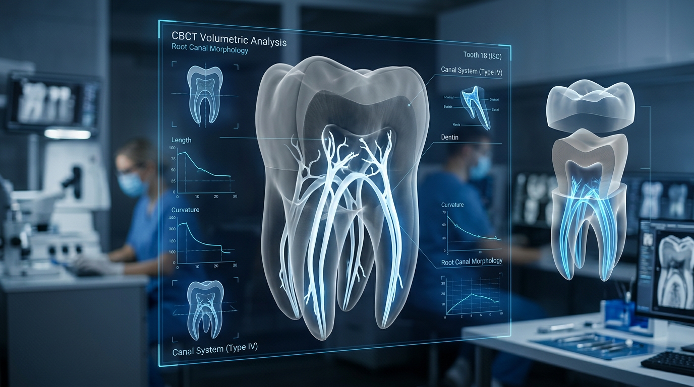
Three dimensional CBCT imaging allows endodontists to study the internal anatomy before treatment begins. Hidden canals, unusual root shapes, and anatomical variations become easier to identify, allowing the clinician to develop a comprehensive treatment plan before starting the procedure.
This level of preparation contributes to more efficient treatment and supports long term tooth preservation.
Diagnosing Cracked Teeth
Cracked teeth can present some of the most difficult diagnostic challenges in dentistry. Patients may experience discomfort while chewing, sensitivity to hot or cold temperatures, or intermittent pain that seems difficult to explain.
Some cracks are extremely small and cannot always be seen during a routine clinical examination. Although not every fracture is directly visible on a CBCT scan, three dimensional imaging often reveals secondary signs that help support diagnosis.
Changes in the surrounding bone, inflammation around the root, or subtle structural irregularities may provide valuable diagnostic information when combined with the clinical examination and other diagnostic tests.
This comprehensive evaluation allows the endodontist to determine the most appropriate course of treatment while helping patients better understand the condition affecting their tooth.
Evaluating Dental Infections
Infections affecting the dental pulp often begin deep inside the tooth before extending into the surrounding tissues. During the early stages, these changes may not always be apparent on traditional dental X-rays.
Cone Beam Computed Tomography allows endodontists to evaluate the precise location and extent of infection in three dimensions. Rather than viewing only a single flat image, clinicians can assess the affected tooth from multiple angles, making it easier to identify areas of bone loss, inflammation, or other changes associated with endodontic disease.
This additional diagnostic information supports more accurate treatment planning and provides a clearer understanding of the overall condition before treatment begins.
Planning Complex Root Canal Treatment
Every successful endodontic procedure begins long before treatment starts. Careful planning allows the endodontist to understand the tooth’s anatomy, identify potential challenges, and determine the most appropriate treatment approach. Cone Beam Computed Tomography (CBCT) plays an important role in this process by providing detailed three dimensional images that reveal information not always visible on conventional dental X-rays.
Before treatment begins, CBCT imaging can help evaluate:
- The number of root canals within a tooth
- The length and curvature of each canal
- Areas of canal calcification or narrowing
- Previous dental restorations
- Existing root canal fillings
- Surrounding bone quality
- The location of nearby nerves and anatomical structures
Having this information in advance allows the endodontist to approach treatment with greater precision and confidence. Rather than encountering unexpected anatomical variations during the procedure, many complexities can be identified beforehand, contributing to more efficient treatment and helping preserve as much healthy tooth structure as possible.
CBCT Versus Traditional Dental X-rays
Many patients wonder whether Cone Beam Computed Tomography replaces traditional dental X-rays. The answer is no. Both technologies serve valuable purposes, and each plays an important role in modern dental care.
Traditional digital dental X-rays remain the standard for many routine examinations because they provide clear diagnostic information while requiring minimal imaging. They are often sufficient for routine dental evaluations and many common treatment needs.
CBCT, however, is designed for situations where additional detail can improve diagnosis or treatment planning. Rather than producing a single flat image, it creates a complete three dimensional view of the area being examined.
The differences between the two technologies include:
- Traditional dental X-rays produce two dimensional images.
- CBCT produces highly detailed three dimensional images.
- Traditional X-rays may have overlapping anatomical structures.
- CBCT allows each structure to be viewed independently from multiple angles.
- Traditional X-rays provide limited depth information.
- CBCT allows clinicians to evaluate depth, width, and height simultaneously.
- Traditional X-rays are ideal for many routine situations.
- CBCT is especially valuable for complex diagnostic and endodontic cases.
Choosing the appropriate imaging method depends on each patient’s clinical situation. An endodontist will recommend CBCT when the additional diagnostic information provides meaningful benefits for diagnosis or treatment planning.
Benefits of Cone Beam Computed Tomography for Patients
While CBCT is an advanced technology for clinicians, its greatest advantages are experienced by patients. Better imaging leads to better understanding, more precise treatment planning, and greater confidence throughout the treatment process.
Some of the key benefits include:
More Accurate Diagnosis
Because CBCT provides a complete three dimensional view of the tooth and surrounding structures, conditions that might otherwise remain hidden can often be identified more accurately. Improved diagnosis forms the foundation for effective treatment.
Earlier Detection of Complex Problems
Certain dental conditions, including hidden infections, resorption, unusual anatomy, or bone changes, may be identified earlier through three dimensional imaging. Early diagnosis allows treatment decisions to be based on more complete clinical information.
Improved Treatment Planning
Every tooth presents unique anatomical characteristics. CBCT allows the endodontist to evaluate these differences before treatment begins, reducing uncertainty and helping develop an individualized treatment plan.
Better Patient Education
One of the most valuable aspects of CBCT is its ability to help patients understand their diagnosis. Rather than relying solely on verbal explanations, patients can view three dimensional images of their own teeth while the endodontist explains the condition and recommended treatment.
This visual approach often makes complex dental conditions much easier to understand and encourages informed decision making.
Supporting Natural Tooth Preservation
One of the primary goals of endodontics is preserving natural teeth whenever possible. By providing more detailed diagnostic information, CBCT supports conservative treatment planning and helps clinicians make evidence based decisions that protect healthy tooth structure.
When Is a CBCT Scan Recommended?
Traditional dental radiographs remain appropriate for many routine situations. However, there are circumstances in which three dimensional imaging provides important additional information that supports diagnosis and treatment planning.
An endodontist may recommend a CBCT scan in situations such as:
- Persistent tooth pain without a clear cause
- Complex root canal anatomy
- Suspected hidden root canals
- Cracked or fractured teeth
- Dental trauma
- Endodontic retreatment
- Evaluation of persistent infection
- Internal or external root resorption
- Surgical treatment planning
- Assessment of surrounding bone before treatment
Each recommendation is based on the individual clinical situation. The decision to obtain a CBCT scan is made only when the expected diagnostic benefits outweigh the need for additional imaging.
Is Cone Beam Computed Tomography Safe?
Patient safety remains a priority in every aspect of dental care, including diagnostic imaging. Modern CBCT systems are designed specifically for dental applications and use significantly less radiation than conventional medical CT scanners.
Dental professionals carefully follow established guidelines to ensure imaging is performed only when clinically appropriate. The goal is always to obtain the diagnostic information necessary while minimizing radiation exposure.
CBCT technology has continued to evolve, with modern equipment providing higher image quality while using lower radiation doses than earlier generations of scanners. Endodontists also select the smallest appropriate field of view whenever possible, allowing imaging to focus only on the area requiring evaluation.
For patients, this means CBCT provides valuable diagnostic information while maintaining a strong commitment to safety and responsible imaging practices.
Cone Beam Computed Tomography and Other Modern Endodontic Technologies
CBCT is one of several advanced technologies that have improved the quality and precision of modern endodontic care. When combined with other innovations, it allows endodontists to diagnose and treat even complex cases with greater confidence.
Dental Operating Microscopes
Magnification has become an essential part of modern endodontics. Dental operating microscopes allow clinicians to visualize tiny anatomical structures that would otherwise be difficult to see with the naked eye.
When combined with CBCT imaging, the endodontist can first identify complex anatomy on the scan and then use magnification to locate and treat those structures during the procedure.
Digital Radiography
Digital radiographs continue to play an important role before, during, and after treatment. They provide immediate images while reducing processing time and supporting efficient patient care.
Together, digital radiography and CBCT offer complementary diagnostic information that supports accurate clinical decision making.
Electronic Apex Locators
Determining the precise working length of a root canal is essential during treatment. Electronic apex locators help measure the canal accurately, allowing the endodontist to clean and shape the canal while preserving surrounding tissues.
Nickel Titanium Rotary Instruments
Modern nickel titanium instruments have transformed root canal treatment by improving flexibility and efficiency. Their ability to follow natural canal curvature helps preserve tooth structure while supporting thorough cleaning of the canal system.
Ultrasonic Technology
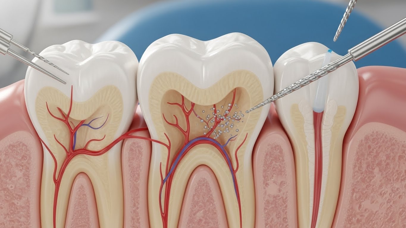
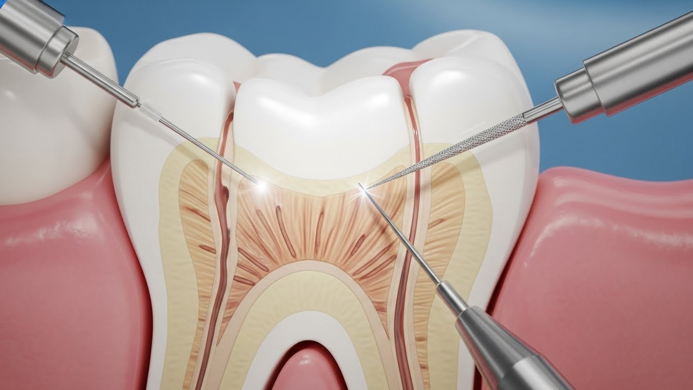
Ultrasonic instruments are frequently used during endodontic procedures to improve visibility, remove previous filling materials when necessary, and assist in locating complex canal anatomy. Combined with detailed CBCT imaging, these technologies contribute to precise and conservative treatment.
The Future of Technology in Endodontics
Advancements in endodontics continue to improve both diagnosis and patient care. Innovations in imaging, digital planning, artificial intelligence, and enhanced visualization are helping specialists evaluate dental conditions with greater accuracy than ever before.
Cone Beam Computed Tomography has become one of the most important developments in this evolution. Its ability to provide highly detailed three dimensional information supports more confident diagnosis, individualized treatment planning, and improved communication between clinicians and patients.
As technology continues to advance, the focus remains unchanged: preserving natural teeth, improving patient outcomes, and delivering high quality endodontic care through evidence based clinical decision making.
Conclusion
Cone Beam Computed Tomography has fundamentally changed the way endodontists diagnose and treat complex dental conditions. By providing detailed three dimensional visualization of teeth, root canals, surrounding bone, and adjacent anatomical structures, CBCT supports more accurate diagnosis, thoughtful treatment planning, and a greater understanding of each patient’s unique dental anatomy.
While conventional dental X-rays remain an important part of everyday dentistry, CBCT offers an additional level of precision when evaluating complex endodontic cases. Whether identifying hidden canals, assessing dental infections, planning retreatment, or evaluating traumatic injuries, this advanced technology provides information that helps clinicians make informed decisions while preserving natural teeth whenever possible.
At Delta Root Canal, modern technology is combined with clinical expertise and patient centered care to support accurate diagnosis and individualized treatment planning. Through the thoughtful use of advanced imaging such as Cone Beam Computed Tomography, patients can feel confident that every treatment recommendation is based on a comprehensive understanding of their oral health and long term dental well-being.
Frequently Asked Questions
What is Cone Beam Computed Tomography (CBCT)?
Summary: Cone Beam Computed Tomography (CBCT) is an advanced three dimensional dental imaging technology used to evaluate teeth, root canals, and surrounding oral structures with greater detail than traditional dental X-rays.
Full Answer: Cone Beam Computed Tomography (CBCT) is a specialized imaging system that creates highly detailed three dimensional images of the teeth, jawbone, root canal system, and surrounding anatomical structures. Endodontists use CBCT to diagnose complex dental conditions, identify hidden canals, evaluate infections, and develop precise treatment plans. The additional level of detail helps improve diagnostic accuracy and supports more predictable treatment outcomes.
Why is CBCT used before root canal treatment?
Summary: CBCT provides detailed three dimensional information that helps endodontists diagnose complex conditions and plan treatment more accurately.
Full Answer: A CBCT scan may be recommended before root canal treatment when additional diagnostic information is needed. Three dimensional imaging allows the endodontist to evaluate the number and shape of root canals, identify hidden anatomy, assess surrounding bone, and detect conditions that may not be visible on conventional dental X-rays. This information supports individualized treatment planning and helps preserve as much healthy tooth structure as possible.
Is Cone Beam Computed Tomography better than a traditional dental X-ray?
Summary: Both imaging methods are valuable, but CBCT provides significantly more detail for complex endodontic cases.
Full Answer: Traditional dental X-rays remain an important part of routine dental care and are appropriate for many examinations. CBCT is used when additional diagnostic detail is beneficial. Because it produces a three dimensional image instead of a flat two dimensional view, CBCT allows endodontists to evaluate complex anatomy, bone, root canals, and surrounding tissues with greater accuracy.
Can a CBCT scan detect hidden root canals?
Summary: Yes. CBCT imaging often helps identify additional or unusually shaped root canals that may not be visible on conventional X-rays.
Full Answer: Every tooth has unique anatomy, and some teeth contain extra canals or complex canal systems. Cone Beam Computed Tomography provides a three dimensional view that helps endodontists identify these anatomical variations before treatment begins. Detecting hidden canals supports comprehensive root canal therapy and contributes to long term treatment success.
Can CBCT help diagnose cracked teeth?
Summary: CBCT can provide valuable diagnostic information when evaluating certain cracked teeth and related conditions.
Full Answer: While very small cracks may not always be directly visible, CBCT imaging often reveals surrounding bone changes, inflammation, fracture patterns, or other findings that assist the endodontist during diagnosis. When combined with a clinical examination and magnification, CBCT becomes an important part of evaluating teeth with suspected cracks.
Is a CBCT scan safe?
Summary: Modern dental CBCT systems are designed to provide detailed diagnostic information while following established radiation safety guidelines.
Full Answer: Dental professionals recommend CBCT only when it is clinically appropriate. Modern CBCT units are designed specifically for dental imaging and typically expose patients to significantly less radiation than conventional medical CT scanners. Endodontists also use the smallest imaging area necessary to obtain the required diagnostic information while prioritizing patient safety.
How long does a CBCT scan take?
Summary: Most CBCT scans are completed in less than one minute.
Full Answer: The imaging portion of a Cone Beam Computed Tomography scan is very quick and usually takes less than one minute. The overall appointment may require only a few additional minutes while the images are processed and reviewed. Patients typically remain seated or standing comfortably throughout the scanning process.
Why is a CBCT scan part of every patient’s evaluation?
Summary: We routinely use CBCT imaging because it provides detailed three-dimensional information that supports accurate diagnosis and treatment planning.
Full Answer: A CBCT scan gives the endodontist a detailed three-dimensional view of the tooth, its roots, surrounding bone, and nearby structures. This helps identify complex anatomy, infections, fractures, resorption, and other concerns that may not be clearly visible on standard dental X-rays. In our practice, CBCT imaging is a routine part of every patient’s evaluation, helping us plan treatment with greater precision and confidence.
What are the benefits of Cone Beam Computed Tomography for patients?
Summary: CBCT improves diagnosis, treatment planning, patient education, and overall clinical precision.
Full Answer: Cone Beam Computed Tomography offers several important benefits, including improved visualization of tooth anatomy, more accurate diagnosis of complex dental conditions, individualized treatment planning, better communication between the patient and endodontist, and support for preserving natural teeth whenever possible. The technology allows treatment decisions to be based on highly detailed clinical information.
Can CBCT improve the success of root canal treatment?
Summary: CBCT supports successful treatment by helping endodontists make more informed clinical decisions before treatment begins.
Full Answer: Although no technology can guarantee treatment outcomes, Cone Beam Computed Tomography contributes to more accurate diagnosis and comprehensive treatment planning. By identifying hidden anatomy, infections, fractures, and other factors before treatment, CBCT helps endodontists perform procedures with greater confidence and precision.
Why is advanced technology important in modern endodontics?
Summary: Advanced technology allows endodontists to diagnose conditions more accurately while providing efficient, patient centered care.
Full Answer: Modern endodontic technologies, including Cone Beam Computed Tomography, operating microscopes, digital radiography, electronic apex locators, and nickel titanium rotary instruments, have significantly improved the diagnosis and treatment of complex dental conditions. These innovations help clinicians preserve natural teeth, improve treatment accuracy, and provide a more comfortable patient experience.

 Improves Modern Endodontic Treatment)
 Improves Modern Endodontic Treatment&description=&image=https://deltarootcanal.com/wp-content/uploads/2026/08/Cone-Beam-Computed-Tomography-CBCT-in-Endodontics2-1024x572.jpeg)